The vagus nerve is one of the biggest cranial nerves. It interconnects the brain with almost all internal organs, including the heart, the respiratory tract, the kidney, and the digestive tract. It mediates important reflexes for autonomic functions and modulates a wide range of body functions, making it an attractive target for bioelectronic interventions. On the flip side, though, this many-sidedness carries the risk of causing unwanted side effects. To activate only those fibers that affect the desired target function, precise information on the structural organization of the nerve is needed.
A large group of scientists at several research institutions headed by the Feinstein Institutes for Medical Research has now undertaken the painstaking effort to make a detailed functional map the fascicles in the vagus nerve and the organs and functions they relate to.
To this end, they used pigs as an animal model. In pigs, the vagus nerve is very similar to humans as far as its structural dimensions and functional organization are concerned. With anatomical methods, the authors meticulously traced the 40-50 fascicles of the vagus in the neck area back to their end organs, revealing organ-specific clusters.
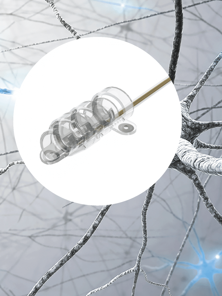
To determine the functional significance of these clusters, they used a unique multi-contact cuff electrode with a helical design that CorTec custom-made for their approach. The cuff has 8 contacts, distributed over the length of the electrode. The cuff is pre-shaped such that it wraps around the nerve in a helical fashion. It is secured in place by 2 outer support loops. In the final configuration, the 8 contacts are uniformly arranged around the nerve diameter, making it possible to stimulate the nerve from 8 different directions. Each contact specifically activates the nerve sector above which it is situated.
The results that the authors obtained with this approach were astonishing: Keeping the electrode at one and the same position, stimulating through the individual contacts could elicit very different functional responses. While one contact would elicit changes in heart rate, for example, the neighboring one could, for instance, affect blood pressure, breathing rate or laryngeal muscle contractions. This impressively shows how profoundly important it is to know and control exactly where you are stimulating around a nerve. Precise positioning and spatial resolution of neuromodulation devices is key to designing specific bioelectronic therapies that produce the desired outcomes and efficiently avoid side-effects.
Citation:
Naveen Jayaprakash, Viktor Toth, Weiguo Song, Avantika Vardhan, Todd Levy, Jacquelyn Tomaio, Khaled Qanud, Ibrahim Mughrabi, Yiela Saperstein, Yao-Chuan Chang, Moontahinaz Rob, Anna Daytz, Adam Abbas, Jason Ashville, Anna Vikatos, Umair Ahmed, Anil Vegesna, Zeinab Nassrallah, Bruce T. Volpe, Kevin J. Tracey, Yousef Al-Abed, Timir Datta-Chaudhuri, Larry Miller, Mary F. Barbe, Sunhee C. Lee, Theodoros P. Zanos, Stavros Zanos
Organ- and function-specific anatomical organization and bioelectronic modulation of the vagus nerve
bioRxiv 2022.03.07.483266; doi: https://doi.org/10.1101/2022.03.07.483266
Note: This article is a preprint and has not been certified by peer review
